Figure 8
COVID-19 New variant and air pollution relationship: how airborne mutagens agent can act on genoma viruses expression: Hypothesis of work
Luisetto M*, Naseer Almukthar, Gamal Abdul Hamid, Tarro G, Khaled Edbey, Nili BA, Ghulam Rasool Mashori, Ahmed Yesvi Rafa and Latyshev O Yu
Published: 16 February, 2021 | Volume 5 - Issue 1 | Pages: 022-031
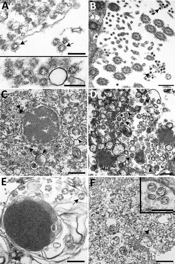
Figure 8:
Ultrastructural- features of severe acute respiratory syndrome – SARS coronavirus 2 lung- infection in fatal coronavirus disease. A) Top: alveolar space containing extracellular virions (arrows) with prominent surface projections. Bottom: cluster of virions in the alveolar- space ( A. S.). Scale bars indicate 200 nm. B) Extracellular virions (arrow) associated with ciliated cells of the upper airway. Scale bar indicates 200 nm. C) Membrane-bound vacuoles (arrows) containing viral- particles within the cytoplasm of an infected type II pneumocyte; surfactant (lamellated -material) indicted by arrowheads. Scale bar indicates 1 μm. D) Membrane-bound vacuole (double-headed arrow in panel C) containing virus -particles (arrows) with the characteristic black dots that are cross-sections through the viral nucleocapsid. Arrowheads indicate vacuolar- membrane. Scale bar indicates 200 nm. E) Viral particles (arrow) within a phagosome of an alveolar macrophage. Scale bar: 200 nm. F) Viral particles within a portion of a hyaline- membrane. Scale bar indicates 800 nm. Inset: Higher magnification of virus- particles indicated by arrow; scale bar indicates 200 nm. Image from Vol-26, Number 9—September 2020 Synopsis.
Read Full Article HTML DOI: 10.29328/journal.ijcv.1001031 Cite this Article Read Full Article PDF
More Images
Similar Articles
-
COVID-19 New variant and air pollution relationship: how airborne mutagens agent can act on genoma viruses expression: Hypothesis of workLuisetto M*,Naseer Almukthar,Gamal Abdul Hamid,Tarro G,Khaled Edbey,Nili BA,Ghulam Rasool Mashori,Ahmed Yesvi Rafa,Latyshev O Yu. COVID-19 New variant and air pollution relationship: how airborne mutagens agent can act on genoma viruses expression: Hypothesis of work. . 2021 doi: 10.29328/journal.ijcv.1001031; 5: 022-031
Recently Viewed
-
Management and use of Ash in Britain from the Prehistoric to the Present: Some implications for its PreservationJim Pratt*. Management and use of Ash in Britain from the Prehistoric to the Present: Some implications for its Preservation. Ann Civil Environ Eng. 2024: doi: 10.29328/journal.acee.1001059; 8: 001-011
-
Isolation and Influence of Carbon Source on the Production of Extracellular Polymeric Substance by Bacteria for the Bioremediation of Heavy Metals in Santo Amaro CityLeila Thaise Santana de Oliveira Santos*, Kayque Frota Sampaio, Elisa Esposito, Elinalva Maciel Paulo, Aristóteles Góes-Neto, Amanda da Silva Souza, Taise Bomfim de Jesus. Isolation and Influence of Carbon Source on the Production of Extracellular Polymeric Substance by Bacteria for the Bioremediation of Heavy Metals in Santo Amaro City. Ann Civil Environ Eng. 2024: doi: 10.29328/journal.acee.1001060; 8: 012-017
-
Drinking-water Quality Assessment in Selective Schools from the Mount LebanonWalaa Diab, Mona Farhat, Marwa Rammal, Chaden Moussa Haidar*, Ali Yaacoub, Alaa Hamzeh. Drinking-water Quality Assessment in Selective Schools from the Mount Lebanon. Ann Civil Environ Eng. 2024: doi: 10.29328/journal.acee.1001061; 8: 018-024
-
Evaluation of Soil Water Characteristic Curves of Boron added Sand-bentonite Mixtures using the Evaporation TechniqueSukran Gizem Alpaydin*, Yeliz Yukselen-Aksoy. Evaluation of Soil Water Characteristic Curves of Boron added Sand-bentonite Mixtures using the Evaporation Technique. Ann Civil Environ Eng. 2024: doi: 10.29328/journal.acee.1001062; 8: 025-031
-
Accidents at Waste Storage Facilities: Methods of Struggle with the Consequences of AccidentsE Argal*. Accidents at Waste Storage Facilities: Methods of Struggle with the Consequences of Accidents. Ann Civil Environ Eng. 2024: doi: 10.29328/journal.acee.1001063; 8: 032-038
Most Viewed
-
Evaluation of Biostimulants Based on Recovered Protein Hydrolysates from Animal By-products as Plant Growth EnhancersH Pérez-Aguilar*, M Lacruz-Asaro, F Arán-Ais. Evaluation of Biostimulants Based on Recovered Protein Hydrolysates from Animal By-products as Plant Growth Enhancers. J Plant Sci Phytopathol. 2023 doi: 10.29328/journal.jpsp.1001104; 7: 042-047
-
Sinonasal Myxoma Extending into the Orbit in a 4-Year Old: A Case PresentationJulian A Purrinos*, Ramzi Younis. Sinonasal Myxoma Extending into the Orbit in a 4-Year Old: A Case Presentation. Arch Case Rep. 2024 doi: 10.29328/journal.acr.1001099; 8: 075-077
-
Feasibility study of magnetic sensing for detecting single-neuron action potentialsDenis Tonini,Kai Wu,Renata Saha,Jian-Ping Wang*. Feasibility study of magnetic sensing for detecting single-neuron action potentials. Ann Biomed Sci Eng. 2022 doi: 10.29328/journal.abse.1001018; 6: 019-029
-
Pediatric Dysgerminoma: Unveiling a Rare Ovarian TumorFaten Limaiem*, Khalil Saffar, Ahmed Halouani. Pediatric Dysgerminoma: Unveiling a Rare Ovarian Tumor. Arch Case Rep. 2024 doi: 10.29328/journal.acr.1001087; 8: 010-013
-
Physical activity can change the physiological and psychological circumstances during COVID-19 pandemic: A narrative reviewKhashayar Maroufi*. Physical activity can change the physiological and psychological circumstances during COVID-19 pandemic: A narrative review. J Sports Med Ther. 2021 doi: 10.29328/journal.jsmt.1001051; 6: 001-007

HSPI: We're glad you're here. Please click "create a new Query" if you are a new visitor to our website and need further information from us.
If you are already a member of our network and need to keep track of any developments regarding a question you have already submitted, click "take me to my Query."


